 Heratsi Branch
 Heratsi Branch
 Heratsi Branch
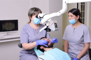
As one of the founders of dental microscope application said: “We can cure what we see”. Yes, for making great progress we need such a tool that will enable us to see what is not seen with the naked eye.
The use of microscope makes the treatment’s outcome predictable and guaranteed.
Thanks to the built-in camera, it is possible to show the patient the true image as well as the entire treatment process, thereby gaining the patient’s confidence and helping him get over his fear.
The microscope has a number of advantages that are important and necessary for the patient.
Due to the peculiarities of the tooth anatomical structure, unfound root canals may cause the appearance of further inflammatory processes and complications. These can lead to a tooth extraction. With the help of the microscope, we can easily detect accessory root canals. The microscope enables more accurate detection of the root canals cracks, as well as the broken endodontic instruments. Years ago, teeth with broken endodontic instruments were subject to extraction. The use of microscope enables to avoid extractions and maintain the high-risk teeth. It enables to carry out complete instrumentation of root canals, treatment and filling under visual control. The dental operating microscope allows us to treat the tooth surface more accurately and precisely. The magnification of tooth surfaces enables precise distinguishing between the healthy and compromised tooth structures thus minimizing the loss of healthy structure. The harmful effect of caries on the healthy tooth structures is a well-known fact.
The treatment of early detected caries guarantees preservation of the teeth.
Subsequently, distinguishing between healthy and compromised dental structures inside the subgingival carious lesions is of great importance. One of the main reasons for dental fractures can be cracks occurring in the enamel and dentin layers as well as inside restorations. Those factors can lead to the disintegrity of the pulp chamber thus causing root canal infections.
With the help of a dental microscope, those cracks can be reviewed in the due time and a proper treatment can be carried out.
While preparing the teeth, the dental microscope helps to avoid soft tissue and mucous membrane damages. In case of surgery, it facilitates implant placement, gingivoplasty and suturing as well as checking the proper fit of the provisionals in prosthodontics in order to maintain the integrity of the tooth thus avoiding any leakage of microbes. The dental microscope provides a magnification up to 40x depending on the procedures taken. The photos and recording taken throughout the procedures can be archived and have a chance of comparing. Finally, the work is carried out in ergonomic working position thus increasing the productivity of the doctor and the nurse and the overall efficiency of the work. The optimal working distance between doctor and the patient is 40-42 cm, which makes the patient feel comfortable, preserving his/her personal space.

